
Digital Dental X-Rays at Mary St Dental Gympie
Embrace the future of dental diagnostics with Low Dose Digital Radiography at Mary St Dental Gympie.
At Mary St Dental in Gympie, we use digital dental radiography to assist with examining teeth and surrounding oral structures.
Digital dental X-rays provide information that may not be visible during a clinical examination, helping dentists investigate particular concerns and plan appropriate treatment.
Dental radiography involves exposure to a small amount of ionising radiation. Our dentists consider each patient’s clinical needs before recommending imaging, with the aim of obtaining necessary diagnostic information while keeping radiation exposure appropriately low.
Not every patient requires dental X-rays at every appointment.
What Is Low-Dose Digital Radiography?
Digital dental radiography uses X-rays and electronic imaging sensors to produce images of teeth, roots and surrounding structures.
These images may assist with identifying dental decay between teeth, assessing infections around tooth roots, examining supporting bone and investigating other dental concerns.
Digital imaging systems offer practical benefits, including electronic image capture, display and storage.
Depending on the equipment and examination settings, some digital systems may use lower radiation doses than older film-based techniques.
However, radiation exposure varies according to the type of examination, equipment and imaging settings. A lower radiation dose does not mean the procedure is completely free of risk.
Your dentist will recommend imaging when the expected diagnostic benefit justifies the exposure.
Why Might Digital Dental X-Rays Be Recommended?
Dental X-rays may be useful when a clinical examination alone does not provide sufficient information about a particular dental concern.
Depending on the imaging technique, X-rays can assist with assessing:
- Decay between teeth or beneath existing restorations.
- Infection or changes around tooth roots.
- Supporting bone levels and certain periodontal conditions.
- Tooth development and eruption.
- The position of impacted or unerupted teeth.
- Particular restorative or surgical treatment requirements.
Different dental X-ray techniques provide different types of information.
For example, bitewing X-rays are commonly used to assess areas between teeth, while periapical images show a tooth, its roots and surrounding structures.
Panoramic imaging (OPG) provides a broader view of the teeth and jaws.
In selected circumstances, 3D cone beam computed tomography (CBCT) may be recommended when additional anatomical information is required, such as for particular dental implant or surgical assessments.
CBCT generally involves greater radiation exposure than conventional dental X-rays and is not required for routine dental examinations.
The type and frequency of imaging depend on individual clinical findings, dental history and risk factors.
What Can You Expect During Digital Radiography?
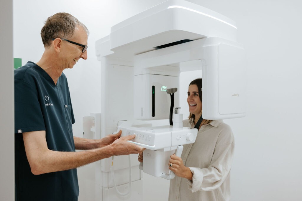
If dental imaging is recommended, your dental practitioner will explain the purpose of the examination and what it involves.
For some X-rays, a small sensor or holder is positioned inside the mouth.
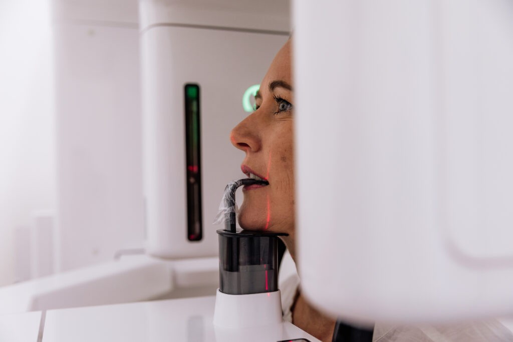
Other examinations use equipment positioned outside the mouth, such as panoramic or CBCT imaging systems.
You may be asked to remain still briefly while the image is captured.
The imaging process is generally quick, although positioning the equipment can cause temporary discomfort or trigger a gag reflex in some patients.
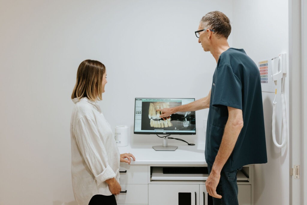
Digital images can usually be viewed electronically, allowing your dentist to discuss relevant findings and explain any recommended treatment options.
If you are pregnant, may be pregnant or have concerns about radiation exposure, please inform your dental practitioner so the clinical need and appropriate precautions can be discussed.
Frequently Asked Questions About Low-Dose Digital Radiography
Dental X-rays generally involve very low levels of ionising radiation, and the associated risks are considered low when imaging is clinically justified and appropriately performed.
However, examinations involving ionising radiation are not entirely free of risk.
Your dentist will assess whether the expected diagnostic benefit justifies the exposure and use appropriate imaging techniques and settings.
Digital radiography uses electronic sensors instead of conventional photographic film to capture dental images.
Digital images can be displayed and stored electronically, and some equipment may achieve suitable diagnostic images using lower radiation doses than older film systems.
The actual radiation dose and image quality depend on the equipment, technique and type of examination.
Your dental practitioner will explain any preparation or precautions appropriate to the examination.
Modern dental radiography uses measures such as appropriate positioning, beam restriction and optimised exposure settings to reduce unnecessary radiation.
Lead aprons are not routinely required for many contemporary dental X-ray procedures, although individual circumstances and clinical protocols may affect their use.
Yes. Dental X-rays may be recommended for children when there is a clinical reason for imaging.
The decision depends on the child’s dental development, oral health, risk of tooth decay and other individual factors.
Exposure settings and techniques should be appropriate for the child’s age and clinical needs.
Not every child requires dental X-rays during every dental examination.
There is no single imaging schedule suitable for everyone.
Your dentist considers your dental history, examination findings, risk of decay, gum health and other clinical factors before recommending X-rays.
Some patients may require imaging more frequently than others, while some appointments may not require any new X-rays.
Discuss Digital Dental Imaging at Mary St Dental Gympie
If you have questions about dental X-rays or the imaging used during dental treatment, our team can provide further information.
Mary St Dental welcomes patients from Gympie, Southside, Monkland, Curra, Glastonbury and surrounding communities.
Your dentist can explain whether imaging is appropriate for your circumstances, the type of examination recommended and its potential benefits and risks.